 Add to Chrome
 Add to Firefox
 Add to Edge
CASED: Curriculum Adaptive Sampling for Extreme Data Imbalance
Jul 27, 2018
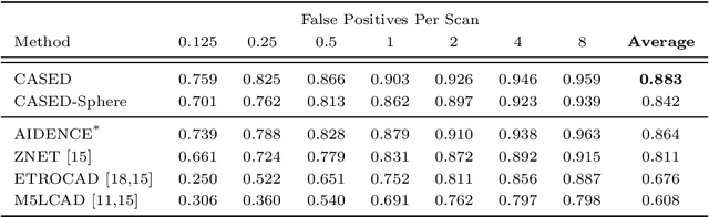
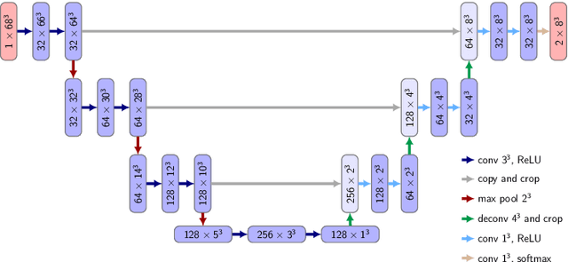
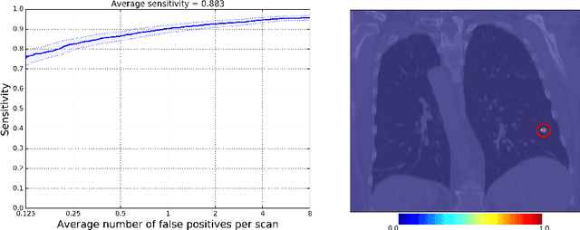
We introduce CASED, a novel curriculum sampling algorithm that facilitates the optimization of deep learning segmentation or detection models on data sets with extreme class imbalance. We evaluate the CASED learning framework on the task of lung nodule detection in chest CT. In contrast to two-stage solutions, wherein nodule candidates are first proposed by a segmentation model and refined by a second detection stage, CASED improves the training of deep nodule segmentation models (e.g. UNet) to the point where state of the art results are achieved using only a trivial detection stage. CASED improves the optimization of deep segmentation models by allowing them to first learn how to distinguish nodules from their immediate surroundings, while continuously adding a greater proportion of difficult-to-classify global context, until uniformly sampling from the empirical data distribution. Using CASED during training yields a minimalist proposal to the lung nodule detection problem that tops the LUNA16 nodule detection benchmark with an average sensitivity score of 88.35%. Furthermore, we find that models trained using CASED are robust to nodule annotation quality by showing that comparable results can be achieved when only a point and radius for each ground truth nodule are provided during training. Finally, the CASED learning framework makes no assumptions with regard to imaging modality or segmentation target and should generalize to other medical imaging problems where class imbalance is a persistent problem.
Deep learning trends for focal brain pathology segmentation in MRI
Jan 24, 2017
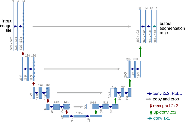
Segmentation of focal (localized) brain pathologies such as brain tumors and brain lesions caused by multiple sclerosis and ischemic strokes are necessary for medical diagnosis, surgical planning and disease development as well as other applications such as tractography. Over the years, attempts have been made to automate this process for both clinical and research reasons. In this regard, machine learning methods have long been a focus of attention. Over the past two years, the medical imaging field has seen a rise in the use of a particular branch of machine learning commonly known as deep learning. In the non-medical computer vision world, deep learning based methods have obtained state-of-the-art results on many datasets. Recent studies in computer aided diagnostics have shown deep learning methods (and especially convolutional neural networks - CNN) to yield promising results. In this chapter, we provide a survey of CNN methods applied to medical imaging with a focus on brain pathology segmentation. In particular, we discuss their characteristic peculiarities and their specific configuration and adjustments that are best suited to segment medical images. We also underline the intrinsic differences deep learning methods have with other machine learning methods.
HeMIS: Hetero-Modal Image Segmentation
Jul 18, 2016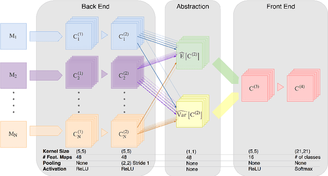
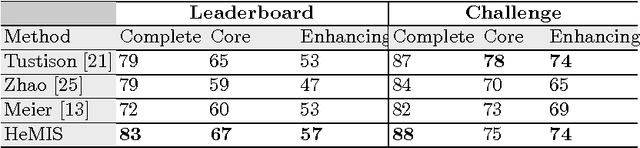
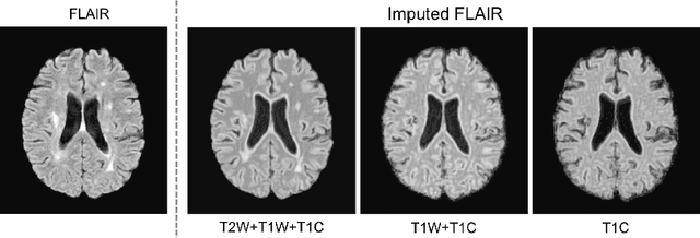
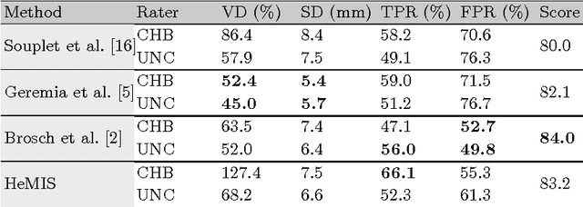
We introduce a deep learning image segmentation framework that is extremely robust to missing imaging modalities. Instead of attempting to impute or synthesize missing data, the proposed approach learns, for each modality, an embedding of the input image into a single latent vector space for which arithmetic operations (such as taking the mean) are well defined. Points in that space, which are averaged over modalities available at inference time, can then be further processed to yield the desired segmentation. As such, any combinatorial subset of available modalities can be provided as input, without having to learn a combinatorial number of imputation models. Evaluated on two neurological MRI datasets (brain tumors and MS lesions), the approach yields state-of-the-art segmentation results when provided with all modalities; moreover, its performance degrades remarkably gracefully when modalities are removed, significantly more so than alternative mean-filling or other synthesis approaches.